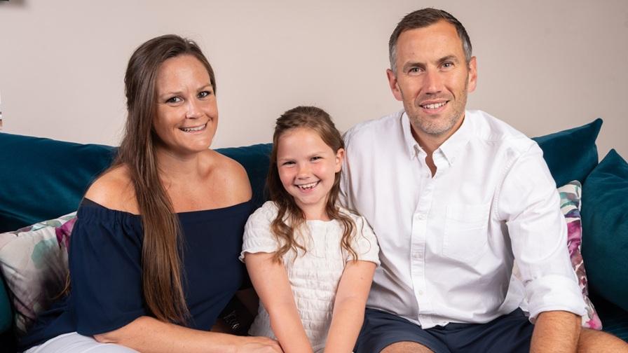

Hattie’s story
Coarctation of the Aorta
Hattie is a very creative and active little girl. “You would never guess that she had been born with a congenital heart condition”, says her mum, Marie.
Marie’s pregnancy had been going smoothly, until the 20-week scan. “We went looking forward to seeing a bit more of the baby,” she remembers. “But everything changed when the sonographer asked us to walk around for a bit, then come back to see if the position of the baby had changed.”
When Marie and husband Sam returned, there was a second, more senior, sonographer in the room. “We soon realised there was more to the ‘walk’,” says Marie. “They said they could only see one main vessel in the heart and one of the chambers looked really small.”
As a former paediatric intensive care nurse, Marie was acutely aware of different heart conditions and feared the worst. “I’d done everything by the book. But you immediately think what have we done wrong?” she says.
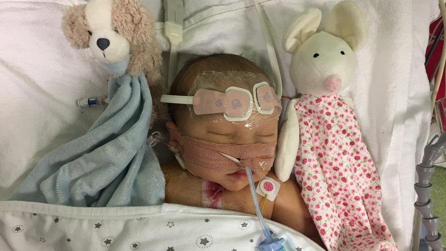
The couple were referred to the Evelina Children’s Hospital in London, where it was confirmed that their baby had severe coarctation of the aorta (CoA). This is a condition in which part of the main artery – the aorta – is narrower than usual, forcing the heart to work harder to pump blood.
CoA affects more than 200 babies born in the UK each year and often requires surgery or other procedures shortly after birth. The condition is not always detected in the womb, putting babies at greater risk of life-threatening complications.
Sadly, some families don't find out there’s a problem until they find themselves facing an emergency in their baby's first days of life”
Knowing Hattie had CoA meant that doctors could monitor her and prepare carefully for her birth.
After ‘a very long and worrying pregnancy’, Marie was induced at 38 weeks. “I got a quick cuddle and then she was taken off to neonatal intensive care with a red hat on,” she recalls.
At 10 days old, Hattie had a successful four-hour operation to repair her heart. “Nothing prepares you enough for seeing your baby attached to a machine, with a breathing tube, wires and drains and a large red dressing on their chest,” says Marie. “But she was soundly sleeping and the medical team were happy.”
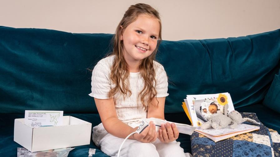
Today, Hattie only needs check-ups every two years. “Her start in life was different to what we expected, but we now see it as a positive. She is an absolute fighter and is fully aware of that time and how special she is.”
Supported by funding from Action Medical Research, Professor Pablo Lamata and his team at King’s College London aim to improve early diagnosis of coarctation of the aorta. They are developing a tool to reconstruct the 3D shape of a baby’s aorta from 2D ultrasound scans like those routinely taken at 20 weeks. This could enable improved early detection – reducing uncertainty for families and improving outcomes for more babies like Hattie.
It would be amazing for more detailed scans to be available and for more babies like Hattie to be picked up before birth”